This article is published in collaboration with The Amherst Student.


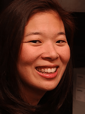
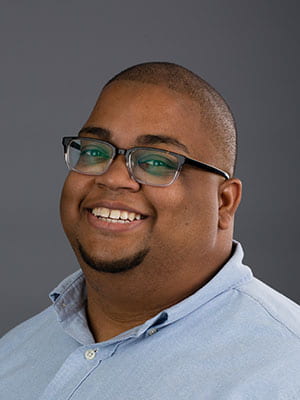
Professors Sally Kim and Marc Edwards of the Biology Department received a Major Research Instrumentation (MRI) grant recently from the National Science Foundation for the acquisition of an integrated Zeiss 980 microscope with Airyscan 2 and Fluorescence Correlation Spectroscopy (FCS) in order to create an advanced microscopy center. We wanted to learn more about the research that won them the award, and how they plan on using this new equipment. Below is the article on our interview with the professors.
First of all congratulations on receiving the award. How would you describe your research interests for someone who isn’t a major?
Prof Edwards: I study cell migrations – how cells make decisions about where and how and why they move. I mainly focus on the phase of the life cycle which involves transitioning from single cells to multicellular (which happens through migration) and back. For the experiment, I use microscopy: both imaging of live cells and stationary cells, to learn more about the defects of movement that we create through mutations in the lab. This grant was a conglomerate of efforts across biology, neuroscience, and chemistry departments. The award we got was a step through building a microscopy center that would facilitate improvements in my research question.
Prof Kim: My research is looking at cellular and molecular mechanisms that explain the synapses, places where neurons communicate. In particular, we’re trying to understand different molecular pathways that are involved with synapse maturation, development, and failure (especially involving autism disorder). So in the lab, we do molecular biology, cell biology, chemistry, and particularly advanced microscopy which requires equipment that we don’t currently have here. Therefore, for this goal, and for the training of future researchers here, whatever they wish to pursue from research to medicine or simply interning in STEM, we applied for this grant and were awarded. Now we’re in the process of hiring a director of microscopy, building an advanced microscopy facility, and we will be teaching a course in the upcoming fall semester on how to operate the equipment. We hope this will be a step towards our vision of where Amherst College can go.
This course you mentioned seems very interesting. Can you talk more about it, to make it more clear for the students who may be interested in taking it?
Prof Kim: Absolutely! It’s called “Quantitative Imaging From Molecules to Cells and Beyond”, Bio/Neuro/BCBP-391. It will be both lecture and lab, taught by Prof. Edwards and me, and the expert we will hire. Our main goal is to train students to become advanced microscopists, teaching them how to operate such equipment to do much more than simply taking pictures. The last part of the course will be students doing a project of their choosing. We hope this course will prepare them for graduate school-level microscopy.
That would help many students who want to go to graduate school after Amherst, so I’m sure the interest level for the registration will be high. Lastly, I wanted to ask about any future real-life applications of your research areas.
Prof. Edwards: For developmental biology, the movement of cells is crucial. It explains how we pass from a single cell, or a group of cells with no particular polarity, to an embryo, a developing organism with patterns that we all share. At the heart of all such developments is the ability of cells to interpret symbols that cue them to move, and to ignore some symbols when appropriate. But sometimes cells do move in ways they shouldn’t, and we know this phenomenon by the name of metastatic cancer. In the end, a lot of how complex organisms function relies on the ability to regulate how cells move from one place to another. The questions that I engage with try to understand how cells process large amounts of information to make such movements and this processing is determined by the details of how cells interact with one another. We want to study these interactions to really understand how this decision-making process takes place. If we could predict and manipulate how cells move, we could prevent many disorders like the type of cancer I mentioned.
Prof. Kim: We’re mainly looking at microstructures where neurons communicate, but we are also interested in how they move. One of the molecules that we study, which I’m also highlighting in one of the courses I teach right now, causes autism when there’s a single-point mutation. We use that as an anchor point to study neurodevelopment. Our end goal is to understand the underlying cellular-molecular mechanisms so that we can propose pathways for certain output functions. Zinc could be used as a potential therapeutic for neurodevelopment, so we do a lot of changing conditions for zinc and synaptic proteins to understand their relations to each other. We believe that this could help us understand autism and find potential pharmacotherapies in the future.
Thank you so much, that was very informative. Your research could help many people in the future, and the new microscopy center is sure to be a highly regarded addition to our Science Center. Thank you for sparing the time to share your thoughts with the Amherst STEM Network!
You must be logged in to post a comment.